1. Introduction
In recent years, the rapid development of virtual reality (VR) technology has provided a revolutionary way for users to interact with the digital world.1 VR has been increasingly used in a variety of fields, including entertainment, education, and therapy, and its effects on users’ cognition and behavior have become a hot topic of research. Particularly in the field of neuroscience and rehabilitation, VR has been used as an innovative tool to demonstrate its potential for brain neurorecovery and cognitive function enhancement.2 As VR technologies become more sophisticated, they can not only simulate complex, realistic environments, but also evoke neurophysiological responses that reflect the physical world. The convergence of technology and neuroscience has opened up new areas for studying the neural correlates of perception, attention, and cognitive processing.3
However, how the level of realism in a VR experience affects the user’s cognitive and neural responses remains an open question. Depending on the display and interaction paradigm, VR systems are commonly categorized as semi-immersive or immersive, distinctions supported by literature on their technological characteristics and user experience.4-5 Semi-immersive VR, often described as a hybrid environment, typically utilizes large projection screens or high-resolution monitors (e.g., desktop VR), allowing users to remain partially aware of their physical surroundings while interacting with digital content via standard interfaces like keyboards or gamepads.4,6 This setup provides a confined but rich virtual experience, frequently used in clinical and training applications.7,8 In contrast, immersive VR is characterized by the use of head-mounted displays (HMDs) that provide a wide field-of-view, head-tracking, and often hand-tracking, creating a strong sensation of being physically present in a simulated environment by occluding the real world and enabling natural, multi-sensory interaction.5,9 The level of immersion is thus closely tied to the technological capabilities for sensory engagement and user agency.10 Immersive VR allows flexible, personalized, and safe multi-modal activities. It operates using natural settings and real-time sensory feedback so that the participants can experience a 360-degree virtual environment, giving them an immersive experience. Immersive VR makes players feel more immersive than semi-immersive VR in a virtual environment.
Semi-immersive VR has been effectively used for a variety of applications, including stroke rehabilitation, post-stroke neurorehabilitation, and pain management for patients with acute or chronic pain.7 For example, one study evaluated the impact of a semi-immersive VR task on the rehabilitation of chronic stroke patients, showing positive outcomes in motor recovery and cognitive examination.8 Additionally, one study compared the use of immersive and semi-immersive VR videos as teaching alternatives in hospitals during the COVID-19 blockade.9 The study utilized a 3D 180° video of the clinical procedures for immersive or non-immersive viewing by students. While the immersive VR group had a higher self-assessment of their expertise, there was no significant difference in learning success between the two groups, suggesting that both immersive and semi-immersive VR have potential for educational purposes. These two different levels of immersion may have different effects on users’ brain activity and cognitive functioning. Therefore, understanding these differences is crucial for designing effective VR applications and treatment programs.
The utility of VR in neuroscience research lies in its ability to create controlled, reproducible, and interactive scenes. This ability is critical to understanding how sensory inputs are integrated and processed and how these processes relate to behavior. Recent advances in neuroimaging techniques, such as functional near-infrared spectroscopy (fNIRS), further enable researchers to examine the neural mechanisms behind VR experiences. In recent years, fNIRS has been exceptionally good for exploring brain function from the data regarding simple blood-oxygen-level changes, by recording and detecting different responses and connectivity between different brain regions when the brain engages in a variety of complex activities. In this experiment, to explore the different effects of immersive VR, semi-immersive VR, and the real environment, fNIRS was used in conjunction to scrutinize the brain activities of the subjects, in order to explore the inducing factors and mechanisms of the different brain modes, with a view to finding more suitable rehabilitation strategies and research results for neurorehabilitation patients.
2. Materials and methods
2.1. Participants
For the study, 30 healthy participants (18 men and 12 women, age: 24.3 ± 3.1 years, education level: all participants had attained at least a bachelor’s degree) participated voluntarily in this experiment. When asked to indicate their dominant hand and with normal or corrected-to-normal vision, all participants reported a right-handedness. None of them had a psychiatric or neurologic clinical history. Participants were aware of the purpose and procedures of the experiment. The study was approved by the Ethics Committee of the Third Affiliated Hospital of Sun Yat-sen University (Approval Number: [2022]02-305-01). All participants provided written informed consent before their participation.
In compliance with ethical guidelines, before beginning the study, a questionnaire for Mini-Mental State Examination (MMSE) was completed by the participants. The MMSE results were obtained immediately so that participants with cognitive impairments could be excluded (subjects with a score above 23). None of the participants were excluded from the study (Mean = 28.83, SD = 1.29).
2.2. Instruments
Two types of instruments (psychological and physiological) were used in this study to measure the participants’ experience during the exposure session.
2.2.1. Psychological questionnaire
2.2.1.1. Online questionnaire
The questionnaire used in this study is the same as the one previously used in previous articles.11 An online questionnaire was administered during the task phase of each reception of VR in order to examine the validity of virtual illusions from a subjective perspective, specifically the illusory sense of body ownership and agency over the virtual body. The online questionnaire was administered verbally by the researcher, and subjects had to answer yes or no to report their level of agreement with the questionnaire statements. The questionnaire consisted of four statements (statement 1 through statement 4 in Table 1), two on the sense of body ownership and two on the sense of agency (one of which was a “real statement” used to check for the actual presence of the phantasmagoria and the other a “control statement”) (Table 1). The questionnaire was administered in the middle of the task each time.
Table 1. Online questionnaire in the virtual reality task.
Online
questionnaire
|
|
Sense of body ownership
|
I feel as if I am looking at my body.
|
Sense of body ownership control
|
I feel as if the virtual body belongs to another person.
|
Sense of agency
|
The virtual body moves just as I want, as if I am controlling it.
|
Sense of agency control
|
I feel as is the virtual body is controlling my will.
|
2.2.1.2. Offline questionnaire
Following the VR tasks in each session, participants were asked to complete another questionnaire containing more detailed statements about subjective feelings of movement, motor control, and physical strength (see Table 2).12,13
Table 2. Offline questionnaire in the virtual reality task.
Offline questionnaire
|
|
Located
|
I felt as if my body was located where I saw the virtual body to be.
|
Sense of ownership
|
I felt that the virtual body was my own body.
|
Standing
|
I felt that I was standing upright.
|
My movements
|
I felt that the leg movements of the virtual body were my movements.
|
Sense of agency
|
I felt that the leg movements of the virtual body were caused by my movements.
|
Sense of ownership control
|
I felt that the virtual body belonged to someone else.
|
Effort
|
I felt I had to give extra physical effort when the virtual body was walking faster.
|
Vection
|
I felt that I was moving through space rather than the world moving past me.
|
Walking
|
I felt that I was walking.
|
Dragged
|
I felt that I was being dragged.
|
Sliding
|
I felt that I was sliding.
|
2.2.2. fNIRS
Hemodynamic changes during the VR task were monitored in real time using a two-wavelength (730 and 850 nm) fNIRS device (Nirsmart, Danyang Huichuang Medical Equipment Co., Ltd., China). Twelve emitters and 10 detectors comprising 24 channels were placed in accordance with the international 10-20 system, using a fixed 3 cm source-detector spacing. Cortical hemodynamic responses were collected and recorded at a sampling rate of 11 Hz.
Statistical parameters from the Montreal Neurological Institute were mapped to the NIRS_SPM software to calculate the coordinates of each source and detector position. The software provides Brodmann regions corresponding to each fNIRS channel. Based on the Brodmann partitioning, six regions of interest were identified in the bilateral prefrontal cortex (PFC), dorsolateral prefrontal cortex (DLPFC), frontal eye fields (FEFs), premotor cortex (PMC), primary somatosensory cortex (S1), primary motor cortex (M1), and occipital cortex (OC) (see Fig. 1).
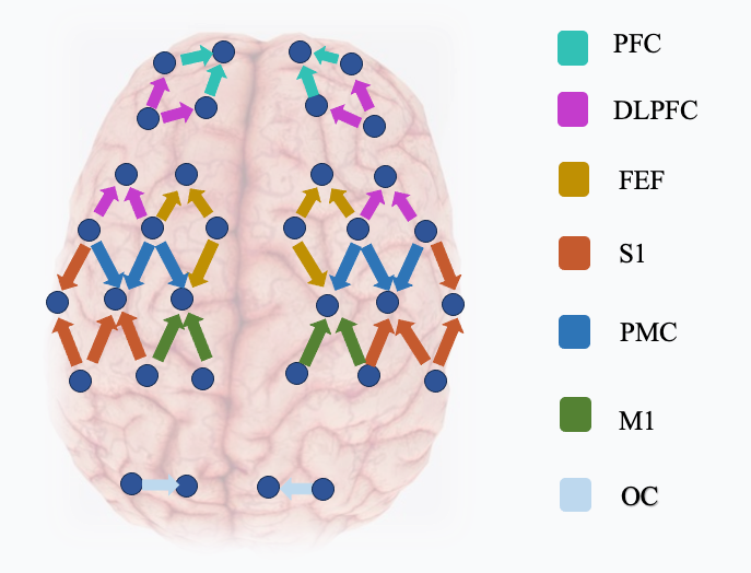 Fig. 1. Schematic diagram of the fNIRS.
 Abbreviations: DLPFC, dorsolateral prefrontal cortex; FEF, frontal eye field; OC, occipital cortex; PFC, prefrontal cortex; PMC, premotor cortex; M1, primary motor cortex; S1, primary somatosensory cortex.
2.3. Experimental design
A randomized, within-subjects (repeated-measures) design was employed for this study. All participants performed all three experimental conditions (immersive VR, semi-immersive VR, and the real environment) in a randomized order to mitigate sequence effects and learning bias. The randomization sequence was generated for each participant individually using a computer-based random number generator (www.random.org, using a simple randomization algorithm). The resulting sequence (e.g., Order A: T1-T3-T2, Order B: T2-T1-T3, etc.) was recorded and stored separately by a research assistant who was not involved in data collection. Participants were assigned to their unique task sequence upon arrival. After signing an informed consent form and receiving written instructions explaining the procedure, participants underwent a familiarization exercise with the different experimental conditions. Each task lasted 10 min (see Fig. 2). Prior to the start of each experimental task, participants were asked to rest for 24 h. During the completion of the tasks, participants were asked to wear a mobile fNIRS to collect brain data, and to answer the appropriate questionnaire during and after completion of the task.
The experimental conditions for each task were as follows: a visual cue “begin” appeared to indicate the start of the experiment. In the semi-immersive and immersive VR tasks, participants were asked to hit the ball with a right-handed grip. The VR program was set up to launch a table tennis ball every 2 s for the participant to hit. The location of the table tennis ball was randomized. If the participant succeeded in hitting the table tennis ball back, the robot would play against the participant until the participant failed to catch the ball. In the real table tennis task, the trained experimenter will launch the ping-pong ball to the participant at a frequency of once every 2 s. If the participant successfully returns the table tennis ball, the experimenter will play against the participant until the participant fails to catch the ball. The VR program and the experimenter playing the role of a ball-firing machine were set to a medium training difficulty, i.e., the participant hit the ball between two and four times in the same round of strikes. This was to prevent the participant from losing interest or patience.
 Fig. 2. Experimental protocol.
 Abbreviation: VR, virtual reality.
2.4. VR equipment
The technical parameters of the HMD (90 Hz refresh rate, 360° tracking) and the large-screen TV setup were aligned with the standard definitions of immersive and semi-immersive VR systems, respectively.5,14
To present immersive VR conditions, a HMD (HTC Vive Pro) with a resolution of 2880 × 1600 pixels (1440 × 1600 pixels per eye) and a refresh rate of 90 Hz was used. This is an immersive 3D VR system widely used in the gaming industry. It can accurately determine the position of the HMD using modern tracking technology to achieve a 360-degree VR stereoscopic presentation from an egocentric perspective.
For the semi-immersive VR condition, a 65-inch TV with a resolution of 2880 × 1600 pixels (1440 × 1600 pixels per eye) and a refresh rate of 90 Hz was used as the monitor. An Xbox was also equipped as the output. Participants stood in front of the TV at a distance of 1.5 meters to complete the corresponding task.
2.5. Data analysis
In this study, the hemodynamic responses were predominantly monitored using the HbO2 signal, favored for its increased sensitivity to local cerebral blood flow compared to HbR. Our established methods for preprocessing fNIRS data, as delineated in previous works,15–18 were rigorously applied to ensure data accuracy and reliability. Initially, the raw absorbance signals obtained via fNIRS were subjected to meticulous filtering. This was achieved using a zero-phase fifth-order Butterworth filter within the frequency range of 0.0095–2 Hz. Such filtering effectively eliminated extraneous noise and attenuated low-frequency drifts, which are commonly observed in raw fNIRS data. This step is crucial to isolate true hemodynamic signals from physiological and instrumental noise. Subsequently, the filtered signals were converted into concentration changes of HbO2 using an advanced application of the Beer–Lambert law, tailored to accommodate the complexities of cerebral tissue optics. This conversion is essential to derive meaningful hemodynamic information from the optical density data. Moreover, to capture the oscillatory signals that correlate with neural activity, we utilized a complex Morlet wavelet transform. This approach focuses on a frequency range of 0.01–0.08 Hz, a bandwidth typically associated with cerebral hemodynamic fluctuations. Automatic outlier detection and correction were also implemented using moving average and cubic spline interpolation techniques. These procedures are instrumental in rectifying sporadic anomalies in the data, ensuring a smoother and more consistent signal for the analysis.
Lastly, the cleaned and processed fNIRS data were subjected to functional connectivity analysis using wavelet phase coherence (WPCO). This technique discerns the phase relationships between signals from different brain regions, with the WPCO value ranging from 0 to 1, indicating the level of phase synchronization. High WPCO values signify strong connectivity, while lower values imply reduced correlation. The analysis was further validated using an amplitude-adaptive Fourier transform technique, contrasting the original signals with a series of surrogate signals to confirm significant phase coherence.
The wavelet transform was used to obtain the phase dynamic information of the oscillating signals.19 The phase dynamic information identifies an effective network model between the 52 channels measured by fNIRS, which is based on the coupling function.20 The parameters describing the coupling model were inferred by dynamic Bayesian inference, revealing the functional rules of interactions in the brain dynamical system.21 The coupling relationship (including coupling strength and direction) between every two channels was quantitatively described by the model parameters. The instantaneous phase and its possible relationship with WPCO were determined.
IBM SPSS Statistics for Windows (version 25.0; IBM Corp, Armonk, NY) was used for statistical analysis. The Shapiro–Wilk test was used to check the normality of the data. Differences in hemodynamic changes relative to baseline and across tasks were compared using repeated-measures analysis of variance (ANOVA). Given the multiple comparisons conducted across the 24 fNIRS channels and several conditions, the false discovery rate (FDR) correction was applied to control for the increased risk of Type I errors. The FDR correction was implemented using the Benjamini–Hochberg procedure across all pairwise post hoc comparisons following significant ANOVA results. For any significant main or interaction effects, post hoc pairwise comparisons were conducted using Tukey’s Honestly Significant Difference test. The threshold for statistical significance was set at a corrected P < 0.05. Furthermore, to complement P-values and assess the magnitude of the observed effects, partial eta-squared (ηp²) was calculated and reported for significant ANOVA results.
2.6. Control of variables
To mitigate the impact of inherent differences in sensory feedback across environments and to ensure that the observed neurophysiological responses were primarily attributable to the level of immersion rather than disparate task mechanics, several key variables were controlled.
2.6.1. Visual feedback
The visual task goal was identical across all conditions: to hit a table tennis ball approaching from the other side of a virtual/real table. The ball launch frequency was strictly standardized to one ball every 2 s in all conditions (VR program and human experimenter). The ball trajectory was randomized in both VR and real tasks to maintain similar levels of visual unpredictability and cognitive demand.
2.6.2. Auditory feedback
While the auditory landscape differed (e.g., sounds of the ball hit in VR vs. real acoustics), the core auditory cue—the sound of the ball being struck—was present in all conditions. Participants were instructed to focus on the visual task, and no additional task-irrelevant auditory stimuli were introduced in the VR environments to minimize distraction disparities.
2.6.3. Motor feedback and task mechanics
The primary motor action—a right-handed gripping motion to swing and hit the ball—was consistent. Participants used a table tennis racket in the real environment and a VR controller held in a similar grip in the virtual environments to simulate the action. The objective (hitting the ball back) and the approximate physical range of motion required were matched as closely as possible. The difficulty level was calibrated to be ‘medium’ in all settings, meaning participants typically hit the ball 2–4 times per rally, standardizing the pacing and physical engagement across conditions.
3. Results
3.1. Questionnaire results
Participants reported that in the online questionnaire, ‘I feel as if I am looking at my body’ and ‘I feel as if the virtual body belongs to another person’ were more recognized in the full immersion VR task (
P=0.0087,
t=2.8284, df=27). Whereas in the offline questionnaire, ‘I felt that the virtual body was my own body.’ would be more recognized in the semi-immersive VR task (see Table 3).
3.2. Differences in cerebral activation
Both immersive VR and semi-immersive VR tasks were shown to be significantly stronger than the resting state or real environment in most brain regions, including bilateral PFC, DLPFC, FEF, M1, S1, and PMC. This suggests that either immersion or semi-immersion VR can bring more obvious activation to the brain. In contrast, in the left OC, all three task states were shown to activate more significantly than the resting state. However, in a two-by-two comparison of the three tasks, no significant difference was shown. Similarly, in the right OC, only the semi-immersive VR task was shown to be significantly stronger than the resting state. This may suggest that there were no significant differences between the three tasks in visually relevant brain performance. At the same time, we found that the semi-immersive task was more significantly activated than the immersive task in most brain regions in both different states of the immersive task (see Table 3).
Table 3. The results of cerebral activation.
Brain
|
Side
|
Rest
|
Task 1
|
Task 2
|
Task 3
|
P Value
|
|
FEF
|
L
|
1.96±1.26
|
6.07±2.83
|
8.51±3.90
|
3.00±3.05
|
a, b, d, e, f
|
|
R
|
1.90±1.20
|
5.33±2.58
|
7.77±3.36
|
2.30±2.20
|
a, b, d, e, f
|
|
DLPFC
|
L
|
2.18±0.84
|
6.08±2.43
|
8.20±3.06
|
2.85±2.82
|
a, b, d, e, f
|
|
R
|
2.46±1.11
|
6.42±2.42
|
8.74±2.86
|
2.75±2.47
|
|
|
M1
|
L
|
2.60±1.79
|
6.85±3.32
|
9.50±4.17
|
2.49±2.18
|
a, b, d, e, f
|
|
R
|
3.40±2.64
|
7.80±3.40
|
10.34±3.73
|
2.78±2.50
|
a, b, d, e, f
|
|
PMC
|
L
|
2.05±1.04
|
5.46±1.82
|
7.68±2.59
|
2.46±2.38
|
a, b, d, e, f
|
|
R
|
3.10±2.16
|
8.66±3.64
|
11.67±4.37
|
3.43±3.40
|
a, b, d, e, f
|
|
S1
|
L
|
2.44±6.23
|
6.23±2.23
|
8.30±2.75
|
2.75±2.86
|
a, b, d, e, f
|
|
R
|
2.97±1.62
|
7.44±3.09
|
9.73±3.57
|
2.91±2.85
|
a, b, d, e, f
|
|
OC
|
L
|
3.36±2.45
|
11.77±10.44
|
16.15±10.68
|
11.38±13.97
|
b
|
|
R
|
4.90±12.54
|
15.77±25.43
|
19.09±25.40
|
13.12±16.28
|
a, b, d, e, f
|
|
PFC
|
L
|
2.78±1.16
|
6.71±2.41
|
9.05±3.23
|
3.04±3.08
|
a, b, d, e, f
|
|
R
|
2.86±1.06
|
7.26±2.56
|
9.58±3.11
|
3.04±2.87
|
a, b, d, e, f
|
Abbreviations: a, Rest vs. Task1,
P < 0.05; b, Rest vs. Task2,
P < 0.05; c, Rest vs. Task3,
P < 0.05; d, Task1 vs. Task2,
P < 0.05; e, Task1 vs. Task3,
P < 0.05; f, Task2 vs. Task3,
P < 0.05; Rest, resting state; Task1, immersive VR; Task2, semi-immersive VR; Task3, real table tennis.
3.3. Differences in functional connectivity
In the comparison of functional connectivity, we separately compared the differences between the three task states across brain regions. The comparison revealed that the functional connectivity between semi-immersive in the left FEF, bilateral OC, and other brain regions was significantly stronger in semi-immersive than in full-immersive VR. This may reflect that semi-immersive VR elicits better connectivity in visually relevant brain regions. Meanwhile, the real ping-pong task was significantly stronger than the fully immersive VR task only in the functional connectivity of LPFC-RPFC (T1: Mean= 0.536, T3: Mean= 0.617;
P = 0.028, ηp² = 0.18) and LOC-ROC (T1: Mean= 0.464, T3: Mean= 0.599;
P = 0.019, ηp² = 0.21). Whereas in the comparison with semi-immersive, the table tennis task only had significantly stronger functional connectivity in LOC-ROC (T2: Mean= 0.488, T3: Mean= 0.599;
P = 0.031, ηp² = 0.17) and RPFC-RDLPFC (T2: Mean= 0.484, T3: Mean= 0.559;
P = 0.041, ηp² = 0.15) (see Fig. 3). This could mean that hitting a ping-pong ball in a real situation requires the mobilization of more cognitively relevant brain regions. However, on the other hand, both fully immersive and semi-immersive VR had similar effects on the performance of movement-related brain regions as real-situation table tennis. This may mean that VR can have an equivalent effect on the functional connectivity of brain motor areas as the real environment.
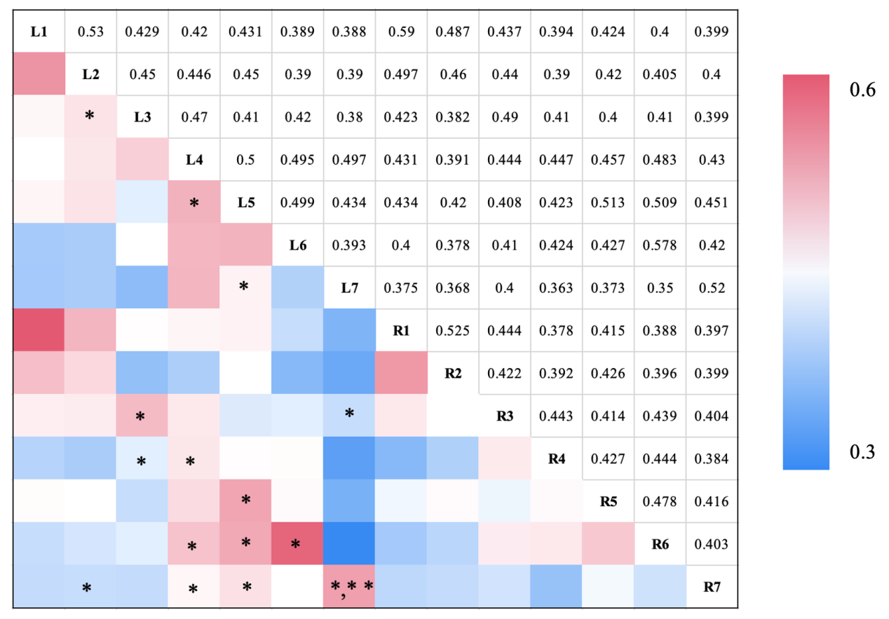 Fig. 3. Significant differences in functional connectivity between experimental conditions.
 This matrix illustrates the pairwise comparisons of functional connectivity between experimental conditions (fully immersive VR task, semi-immersive VR task, and real task). Each cell in the matrix represents a pair of brain regions (the intersection of a row and a column). Significant differences (
P < 0.05, FDR-corrected) in wavelet phase coherence (WPCO) values between conditions are denoted by colored symbols: the red upward-pointing triangle (▲) indicates strong functional connectivity for this brain region under the conditions specified in the row; the blue downward-pointing triangle (▼) indicates weak functional connectivity for this brain region under the conditions specified in the row. The blank cell indicates moderate functional connectivity for this brain region under the conditions specified in the row. Abbreviations: L/R 1, left/right prefrontal cortex; L/R 2, left/right dorsolateral prefrontal cortex; L/R 3, left/right frontal eye field; L/R 4, left/right premotor cortex; L/R 5, left/right primary motor cortex; L/R 6, left/right primary somatosensory cortex; L/R 7: left/right occipital cortex; L/R DLPFC, left/right dorsolateral prefrontal cortex; L/R FEF, left/right frontal eye field; L/R M1, left/right primary motor cortex; L/R OC, left/right occipital cortex; L/R PFC, left/right prefrontal cortex; L/R PMC, left/right premotor cortex; L/R S1, left/right primary somatosensory cortex. Connections marked with an asterisk (*) indicate a statistically significant difference after FDR correction for multiple comparisons (
P < 0.05). In the figure, ‘*’ indicates the resting state showing significant differences when comparing the fully immersive task and ‘**’ indicates the resting state showing significant differences when comparing both the real tasks.
The semi-immersion task showed significant enhancement in functional connectivity in brain regions associated with the left and right occipital cortices (L/R OC) when the semi-immersion task was compared to the immersion task (see Fig. 4). This may indicate that the semi-immersion task has more significant advantages in the input of vision. Whereas, when the semi-immersive task was compared with the real task, the real task showed stronger functional connectivity in the LPFC-RPFC (
P = 0.04). Also, the real task showed stronger enhanced functional connectivity between the left S1 and the right PMC, M1, and S1. This may suggest that in the real ping-pong task, because hitting a ping-pong ball is more complex, it requires the body to mobilize more brain regions to work together.
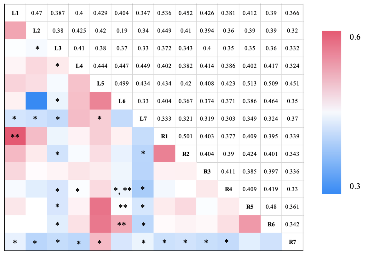 Fig. 4. Significant differences in functional connectivity between the semi-immersion task and immersion task.
 This matrix depicts the wavelet phase coherence (WPCO) values, representing functional connectivity strength, between pairs of cortical regions (rows: R1–R7; columns: L1–L7) under a specific experimental condition. The value within each cell indicates the WPCO coefficient for the corresponding region pair. The color of each cell reflects the magnitude of the coherence, with the gradient from blue to red representing low to high values, as shown in the color bar. Cells are marked with asterisks to denote statistically significant differences (
P < 0.05, FDR-corrected) in WPCO for that specific brain region pair when this condition is compared to others: ‘*’ indicates that the WPCO value for that brain region pair was significantly different between the full-immersion and semi-immersion task conditions. ‘**’ indicates that the WPCO value for that brain region pair was significantly different for both the full-immersion and the real task conditions when each is compared to the remaining condition. Abbreviations: L/R 1: left/right prefrontal cortex (PFC), L/R 2: left/right dorsolateral prefrontal cortex (DLPFC), L/R 3: left/right frontal eye field (FEF), L/R 4: left/right premotor cortex (PMC), L/R 5: left/right primary motor cortex (M1), L/R 6: left/right primary somatosensory cortex (S1), L/R 7: left/right occipital cortex (OC).
Significant differences were found for the real task when comparing the semi-immersive task to the real task in the functional connectivity related to S1 on the right side (PRS1-LS1=0.03, PRS1-LOC=0.04, PRS1-RM1=0.04). Meanwhile, a significant difference in the real task was also found in the functional connectivity of the right PFC and the right DLPFC (
P=0.04) (see Fig. 5). However, from another perspective, both fully immersive and semi-immersive VR had similar effects on the performance of movement-related brain regions and the real situation of ping-pong. This may imply that VR can have equivalent effects to the real environments on the functional connectivity of brain motor regions.
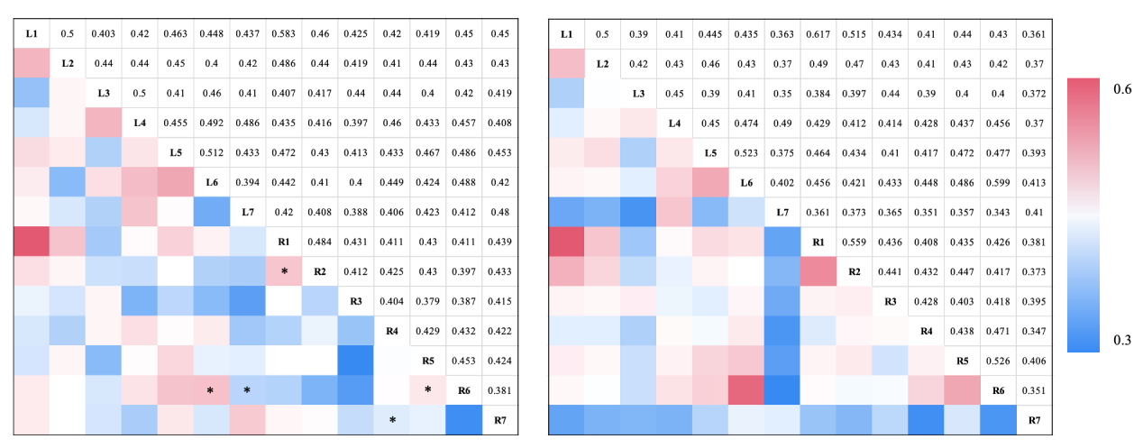 Fig. 5. Comparison of functional connectivity between semi-immersive and real tasks.
 This heatmap matrix illustrates the wavelet phase coherence (WPCO) values, representing the strength of functional connectivity, between pairs of cortical regions (L1–L8 and R1–R7) under the semi-immersive task and the real task conditions. Each cell’s color corresponds to the WPCO value for a specific region pair, with the color gradient from blue to red indicating low to high coherence strength, as defined by the adjacent color bar (range: 0.0 to 0.6). Connections (cells) marked with an asterisk (*) indicate that the functional connectivity (WPCO value) for that specific brain region pair shows a statistically significant difference (
P < 0.05) between the semi-immersive and real task conditions after false discovery rate (FDR) correction for multiple comparisons. Abbreviations: L/R 1, left/right prefrontal cortex; L/R 2, left/right dorsolateral prefrontal cortex; L/R 3, left/right frontal eye field; L/R 4, left/right premotor cortex; L/R 5, left/right primary motor cortex; L/R 6, left/right primary somatosensory cortex; L/R 7: left/right occipital cortex; L1/R1: left/right prefrontal cortex (PFC), L2/R2: left/right dorsolateral prefrontal cortex (DLPFC), L3/R3: left/right frontal eye field (FEF), L4/R4: left/right premotor cortex (PMC), L5/R5: left/right primary motor cortex (M1), L6/R6: left/right primary somatosensory cortex (S1), L7/R7: left/right occipital cortex (OC).
3.4. Differences in effective connectivity
There was more effective connectivity among brain areas in the task states than in the resting state (see Fig. 6). In immersive task, the effective connections were added from R-PMC to L-FEF (
P = 0.03,
F = 5.20), R-PFC (
P = 0.03,
F = 5.03) and R-FEF (
P = 0.04,
F = 4.54). In semi-immersive task, the effective connectivity was added from bilateral-PMC to L-PFC (L:
P = 0.02,
F = 5.45, R:
P = 0.03,
F = 5.20), from R-PMC to L-PMC (
P = 0.01,
F = 8.20), to L-M1 (
P = 0.03,
F = 5.38), and to bilateral-OC (L:
P = 0.02,
F = 7.598, R:
P = 0.02,
F = 6.98).
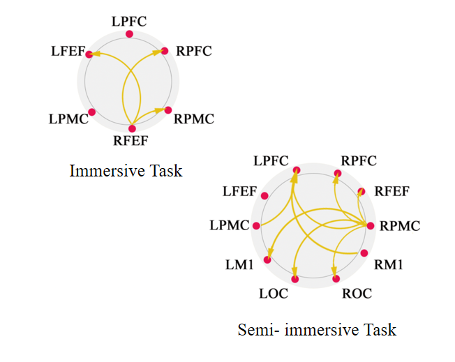 Fig. 6. The effective connectivity graph of the immersive task (left) and semi-immersive task (right).
 Nodes: Red circles represent the regions of interest (ROIs) included in the analysis. Edges: Yellow arrows indicate the presence of effective connectivity (directed functional influence) from one brain region to another, with the arrow direction denoting the direction of information flow. Abbreviations: FEF, frontal eye field; L/R, left/right hemisphere; M1, primary motor cortex; OC, occipital cortex; PFC, prefrontal cortex; PMC, premotor cortex.
4. Discussion
The researchers conducted a thorough investigation into the neurophysiological reactions evoked by immersive VR and semi-immersive VR. The study utilized fNIRS to analyze and map the cortical hemodynamic responses. The study involved a cohort of 30 persons who were in good health. These participants were exposed to various activities in three different settings: immersive VR, semi-immersive VR, and actual environments. Throughout the duration of the tasks, the brain activity of the participants was closely observed and subsequently analyzed.
Significant enhancements in brain activity within the occipital lobes, specifically in areas linked to visual processing, were observed among participants in the immersive VR condition. This implies that the utilization of VR headsets and the accompanying audio-visual stimuli results in increased activation of the visual cortex.10,14 This discovery is consistent with the concept that immersive settings have a greater capacity to capture the user’s visual attention, resulting in a more vibrant and captivating experience.22–24 Differential activation patterns in the brain were seen when comparing immersive and semi-immersive VR situations. The immersive condition predominantly elicited activation in visual regions, but the semi-immersive condition, characterized by less immersive VR experiences (such as viewing a computer screen without a headset), had a more dispersed pattern of brain activation. This finding suggests that situations with lower levels of immersion may necessitate the brain to integrate a greater amount of sensory information from the external, real-world context. Consequently, this integration process results in a more varied pattern of activation throughout various regions of the brain.25–28
It is noteworthy that the study also observed discrepancies in cognitive burden across the two scenarios. The immersive condition predominantly elicited activation in visual regions. In contrast, the semi-immersive condition elicited enhanced functional connectivity within visuospatial networks.29–31 This finding suggests that in semi-immersive setups, where users remain partially aware of the physical surroundings, the brain may engage specific visual and attentional networks to a greater extent to manage and integrate competing inputs from the virtual and real worlds.32–36
The results of the study indicate that the semi-immersive VR condition exhibited notably higher functional connectivity in brain regions associated with visual processing, including the left FEF and the bilateral occipital cortex. This pattern of enhanced functional connectivity, particularly in the left FEF and bilateral OC, suggests that the semi-immersive VR condition might have placed higher demands on visuospatial attention and integration processes compared to the fully immersive condition. A potential explanation for this observation is that in the semi-immersive setup, users remain partially cognizant of their physical surroundings,37 which could require the brain to manage and integrate competing visual inputs from the virtual and real worlds. However, it is important to note that while functional connectivity in visual areas was increased, this does not directly measure ‘cognitive processing’ as a whole. Therefore, we interpret this finding more cautiously as indicative of increased engagement of specific visual and attentional networks, rather than a global increase in cognitive load. Conversely, a significant discovery emerged indicating that tasks performed in real-world settings need greater cognitive involvement, as seen by the increased functional connectivity between the left lateral prefrontal cortex (LPFC) and the right PFC, as well as the bilateral occipital cortex (LOC-ROC). This observation suggests that physical tasks in real-world contexts are characterized by increased complexity and cognitive demands.
The findings indicate that immersive VR has the potential to elicit a robust feeling of presence and ownership of one’s body. However, it appears that semi-immersive VR may elicit a higher level of engagement from the brain’s visual and attentional networks. The potential consequences of this finding could be significant in informing the development of VR systems that are customized to target certain cognitive or therapeutic objectives. For example, situations that necessitate robust visual processing and focused attention may derive advantages from semi-immersive configurations, while those that require a deep sense of presence and embodiment could make use of immersive systems.
The findings derived from the conducted experiments investigating the impact of connectivity have indicated that specific crucial brain regions, specifically those associated with visual processing (such as the lateral occipital cortex and temporal lobe visual areas) and executive functioning (such as the PFC), displayed the increased connectivity during the performance of a semi-immersive VR task in comparison to both a fully immersive VR task and a real-world task. The observed distinction implies that semi-immersive VR has the potential to offer a more equitable approach in stimulating and incorporating sensory information, while mitigating the risk of cognitive overload. This discovery holds clinical significance as it sheds light on the potential of VR technology in enhancing cognitive training and rehabilitation. For instance, in the rehabilitation of unilateral spatial neglect, a condition where patients fail to attend to stimuli on the contralesional side, a semi-immersive setup that requires the brain to integrate competing cues from a virtual task and the residual awareness of the real environment might more effectively engage and train the compromised attentional networks, as suggested by prior work on VR for neglect.37 Conversely, the finding that real-world table tennis engaged prefrontal connectivity more strongly than VR tasks points to the higher cognitive-motor integration demands of natural environments. This underscores a potential application for immersive VR: by designing ecologically valid tasks that progressively increase cognitive load (e.g., complex decision-making in a virtual environment), VR could be used to safely and gradually train these higher-order cognitive and executive functions in patients with frontal lobe impairments or mild cognitive impairment, a direction supported by studies using VR for cognitive assessment and training.28,34 However, a crucial caveat must be emphasized when extrapolating these findings to clinical populations. Our study exclusively involved healthy, young adults. The neurophysiological responses and adaptive patterns observed herein may not directly translate to patients with neurological conditions such as stroke, traumatic brain injury, or neurodegenerative diseases. In patient populations, factors like lesion location, impaired neural connectivity, reduced cognitive reserve, and altered neuroplasticity can fundamentally modulate how the brain responds to VR stimuli.
There are various limitations inherent in our investigation. The limited size of the sample and the absence of diversity may hinder the ability to accurately generalize the findings to the broader community. Furthermore, our participant cohort, while healthy, was relatively homogeneous in terms of age range and was not stratified by factors such as gender or prior VR experience. Individual differences like these are known to potentially influence neurophysiological responses and subjective experiences in VR. For instance, gender differences in spatial navigation or susceptibility to cybersickness, varying levels of VR familiarity affecting the sense of presence, and age-related changes in neural plasticity could all serve as confounding variables that modulate brain activation and connectivity patterns. Our study design did not control for or analyze the effects of these factors, which may have introduced unaccounted variance into our results. Additionally, the specific VR equipment utilized in the study may provide outcomes that are not transferable to alternative systems. This study did not take into consideration individual characteristics, such as prior exposure to VR and personal cognitive strategies, which have the potential to impact neurophysiological responses. Future studies would benefit from a larger, more diverse sample and a design that systematically investigates the role of these individual difference variables. Studies should directly compare the efficacy and neural correlates of immersive versus semi-immersive VR interventions against conventional therapy in specific clinical populations, while carefully controlling for disease severity, stage of recovery, and individual differences in VR tolerance and prior experience. The aforementioned criteria combined indicate a necessity for further detailed research in order to validate and build upon the conclusions that have been provided.
Furthermore, while measures were taken to control task mechanics (e.g., ball frequency, motor action), inherent differences in sensory fidelity (e.g., haptic feedback, visual resolution) between virtual and real environments persist and represent a fundamental characteristic of VR technology itself. These differences may contribute to the variations in brain activation and connectivity observed and should be considered when interpreting the results.
5. Conclusion
This study provides a neurophysiological foundation for tailoring VR systems to specific cognitive or therapeutic objectives. The primary theoretical contribution lies in elucidating the distinct patterns of brain activation and functional connectivity associated with different levels of VR immersion. Specifically, our findings demonstrate that semi-immersive VR elicits a broader pattern of cortical activation and enhances functional connectivity within visuospatial networks (e.g., FEF, OC), suggesting its particular efficacy for applications demanding robust visual attention and processing. Conversely, the finding that real-world tasks engage higher-order cognitive networks (e.g., PFC connectivity) to a greater extent provides a benchmark for understanding the cognitive fidelity of VR simulations.