Background and aims: Diffusion tensor imaging (DTI) has the advantage in revealing subtle pathology of damaged spinal cord more clearly and comprehensively. However, no study to date has elucidated the correlation between motor function recovery and dynamic changes of DTI parameters involving the whole spinal cord after repeated subarachnoid administrations of human umbilical cord mesenchymal stem cells (hUC-MSCs) in canine model of spinal cord injury (SCI). This study aimed to clarify it to quantitatively investigate DTI metrics as potential prognostic indicators for functional recovery of locomotion.
Materials and methods: Eight female beagle canines were subjected to thoracolumbar SCI, and then they received four times of intrathecal transplantation of hUC-MSCs from Week 2 (W2) to W5 (once per week). During the 15-week observation period, motor function of pelvic limb was assessed by using OIby score system at each week. At W1, W8 and W15, DTI scanning rostrally, centrally and caudally within the lesion site was performed and then, multiple radiological parameters, including fractional anisotropy (FA), average diffusion coefficient (ADC), axial diffusivity (AD), radial diffusivity (RD), mean diffusivity (MD), were collected.
Results: Significant improvement of OIby score was observed at the latest ten time points when compared with that at W1. FA rostrally, centrally and caudally within the lesion site revealed the decrease tendency, while all the other radiological indicators at these three regions had the N-shaped trend that showed initial increase and subsequent decrease. Except the comparisons between W15 and W1 regarding cranial MD, cranial RD and caudal RD, the remaining comparisons of radiological data between W15 and W1 and all the DTI metrics between W8 and W1 demonstrated statistical significance. Pearson correlation analysis found that cranial FA (r = 0.723, P = 0.043) at W8, as well as both cranial AD (r = -0.761, P = 0.028) and MD (r = -0.728, P = 0.041) at W15 correlated with the final OIby score. Simple regression model (y = 26.178 - 4.176x, y: OIby score, x: cranial AD value) uncovered a negative linear correlation between rostral AD and locomotion performance both at W15 (R2 = 0.578, β=-0.761, P=0.028).
Conclusions: For the first time, our work demonstrates that in canine model of SCI receiving repeated intrathecal transplantation of hUC-MSCs, cranial FA at 3 weeks post-cytotherapy, as well as both cranial AD and MD at 10 weeks after hUC-MSCs administrations were associated with final regaining of motor function, and rostral AD seems a feasible predictor of behavioral recovery. This study provides DTI clues in predicting sensorimotor function restoration in humans with SCI after repeated subarachnoid transplantation of hUC-MSCs.
Significance Statement
Human umbilical cord mesenchymal stem cells (hUC-MSCs) possesses a promising role in treating spinal cord injury (SCI), a devastating disease of the central nervous system, because of multiple benefits. Diffusion tensor imaging (DTI) is invaluable to capturing microstructural changes distant from the injury epicenter. Based on this advantage, radiological indicators rostrally, centrally and caudally within the damaged spinal cord in canine model were analyzed to make a more comprehensive understanding of pathophysiological change after repeated hUC-MSCs transplantation. For the first time, For the first time, our work demonstrates that in canine model of SCI receiving repeated intrathecal transplantation of hUC-MSCs, cranial FA at 3 weeks post-cytotherapy, as well as both cranial AD and MD at 10 weeks after hUC-MSCs administrations were associated with final regaining of motor function, and rostral AD seems a feasible predictor of behavioral recovery. This study provides DTI clues in predicting sensorimotor function restoration in humans with SCI after repeated subarachnoid transplantation of hUC-MSCs.
Introduction
Spinal cord injury (SCI) is a devastating disease and often leads to irreversible paralysis, sensory deficit, muscle spasm, autonomic disturbance, neuropathic pain, as well as bowel and bladder dysfunction, all of which seriously impact the quality of life 1,2. Up to now, there has been a lack of any effective strategy to reverse the trauma in clinic due to low intrinsic regenerative ability of host neurons 3,4. This has led to a heavy reliance on experimental therapies, including stem cells transplantation. Human umbilical cord mesenchymal stem cells (hUC-MSCs) possess a potential role in treating SCI because of multiple benefits, including easy obtainability, low immunogenicity, fast proliferation, revascularization support, inflammation reduction, multi-lineage differentiation, cellular apoptosis inhibition, remyelination of damaged axons, and production of numerous trophic factors 5-9. Among a variety of cell delivery routes, it has been demonstrated that subarachnoid transplantation has the advantages of more cell engraftment, better tissue sparing, and decreased host immune response, resulting in greater functional recovery of damaged spinal cord 10,11. Because the therapeutic effect of hUC-MSCs transplantation on SCI recovery is dose-dependent 12, repeated cell administrations, which are facilitated by the ease of repeatability from intrathecal infusion, allows for sustained therapeutic effects over time, potentially leading to the maximization of functional restoration after SCI 1,13,14.
Compared with conventional magnetic resonance imaging (MRI) techniques, diffusion tensor imaging (DTI) can reveal the microstructural properties, including orientations, continuity and myelination, in longitudinal neuronal tracts in vivo to assess injury severity both qualitatively and quantitatively. Thus it has great potential for the dynamic evaluation of SCI as a non-invasive imaging approach 15. Due to the heterogeneity between SCI cases regarding injury site, trauma severity, and damage mechanism, it is difficult to accurately investigate the dynamic changes of DTI parameters after repeated subarachnoid administrations of hUC-MSCs in clinic. Considering the resemblance between canine and human in terms of spinal cord size, anatomy, histopathology, and imaging 15,16, canine model of SCI represents a translational alternative to evaluate changes of DTI indicators following aforementioned intervention.
To the best of our knowledge, the correlation between motor function recovery and dynamic changes of DTI parameters involving the whole spinal cord, from the lesion epicenter to rostral and caudal levels, after repeated intrathecal administrations of hUC-MSCs in canine model of SCI is yet to be elucidated. The aim of this study is to clarify it to quantitatively investigate DTI metrics as potential prognostic indicators for locomotion recovery in canine model of SCI and further provides clues for prediction of sensorimotor function restoration in humans suffering from SCI after repeated subarachnoid transplantation of hUC-MSCs.
Materials and methods
Animals
This study was conducted in accordance with the guidelines of the Institutional Animal Care and Use Committee (IACUC) of Sun Yat-Sen University (SYSU) and was approved by this committee (SYSU-IACUC-2018-000130). Eight female beagle canines that were purchased from Nanjing Chaimen Biotechnology Co., Ltd were used in this study. The inclusion criteria were satisfied based on age between 12 and 18 months, body weight between 10 and 12 kg, normal findings after physical and neurological examinations, as well as laboratory blood analysis.
SCI modeling and subarachnoid transplantation
Prior to experimental manipulation, all canines were allowed to acclimatize to the housing facility for a minimum of 1 week. At Week 0 (W0), they were anesthetized as follows: induction by intravenous (IV) propofol (4-5 mg/kg, Peifen, China), followed by isoflurane inhalation for maintenance (minimum alveolar concentration: 1.2-1.8 vol%, Iflurin, China). Oxygen saturation and pulse rate were continuously monitored by using electrocardiography during general anesthesia. A respiration rate of 15 breaths per minute was maintained in a ventilator. After incising the skin, the adipose layer and fascia were cut cautiously and then, paraspinal muscles were bluntly stripped. Hemilaminectomy and ipsilateral facetectomy at T13/L1 level were performed to expose the dural sac with approximate 1-cm long. The spinal cord was compressed ventrally and dorsally by applying an aneurysm clip (Sugita, Japan) with a compression force of 120-140 g for 50 seconds. The lower blade was passed under the dural sac and the upper blade was released rapidly to bruise the spinal cord that showed edema, hyperaemia and hematoma to mimic spinal cord trauma. Following wound irrigation with normal saline, the muscles, fascia and skin were sutured layer by layer. At 12 hours after SCI modeling of canines, drinking and eating could return to normal. Intramuscular injection of vitacoxib (2 mg/kg/d, Orbiepharm, China) was used to reduce pain intensity. Manual micturition and defecation were performed twice daily until their automatic bladder and bowel functions recovered. Moreover, skin nursing care and renewal of waterproof mat in canine cages were carried out once daily to prevent decubitus ulcer.
At W2, after IV sedation consisted of dexmedetomidine (3-5 ug/kg, Orion, Finland) and Zoletil 50 (containing tiletamine and zolazepam, 5-7 mg/kg, Virbac, France), the lumbar puncture at L5/6 level was performed to allow subarachnoid transplantation of hUC-MSCs (1×10⁶ cells/kg) in a 2 ml suspension (supplied by Guangzhou Celera Stem Cell Technology Co., LTD). This was followed by a 2 minutes delay before withdrawal of the injection needle to ensure that all stem cells were delivered within the ducal sac. At W3, each canine underwent a second intrathecal transplantation of hUC-MSCs. Following this pattern, each animal received a third and fourth stem cells infusion at W4 and W5, respectively.
To prevent potential immune rejection upon hUC-MSCs in canines, the immunosuppressant-cyclosporine A (10 mg/kg, Cyonse, China), was administered orally once a day with a stringent 24 hours cycle for the first 3 days after each subarachnoid transplantation of hUC-MSCs, then its dose was reduced to 5 mg/kg in the following 4 days. During the experimental period, all canines were housed in a temperature-controlled (24 ± 2 ℃) room with full access to food and water.
Assessment of pelvic limb locomotion
Motor function of pelvic limb was assessed by using the 15-point OIby score system, which is commonly used to evaluate neurological function in canines with SCI by examining the pain sensation and motor function, including tail movement, weight bearing, gait accuracy and voluntary movement of pelvic limb 17. During this assessment, caninesare allowed to explore in an open field freely for a minimum of 10 steps. Every week, two independent skilled investigators jointly evaluated the animal conditions and then, calculated the OIby score from the start of this experiment till the termination of 15 weeks observation period. The consecutive OIby scores of all canines were used for statistical analysis.
DTI acquisition and processing
DTI scanning was performed at W1, W8 and W15 by using the 3.0 Tesla MRI scanner (Discovery 750, General Electric, United States) under aforementioned IV sedation and assisted ventilation. Each canine was put in supine position and then, placed in the center of a cardiac coil (General Electric, United States). T2 weighted sagittal and axial images were obtained to determine the lesion epicenter and the visible rostral to caudal extent of injured spinal cord by an experienced spine surgeon. Subsequently, DTI sequence scanning was performed. The image post-processing software (Functool) in AW 4.6 workstation (General Electric, United States) was used to reconstruct DTI images in coronal, sagittal and axial planes. The region of interest (ROI) of the lesion epicenter in DTI transverse plane, which excluded cerebrospinal fluid and dura mater as much as possible, was jointly determined by two experienced radiologists. Then, fractional anisotropy (FA), average diffusion coefficient (ADC), axial diffusivity (AD), radial diffusivity (RD), mean diffusivity (MD) were measured in each slice of the lesion epicenter ROI and calculated by the same radiologists jointly. Rostral and caudal ROI were respectively placed within one vertebral body above or below the visible lesion margins, as visualized on T2 weighted sagittal and axial images. By using the same method, all the aforementioned radiological values both rostrally and caudally were obtained (Figure 1). For each animal. the averages values of these imaging metrics rostrally, centrally and caudally within the lesion site at each time point were calculated and used for statistical analysis.
The scan parameters were as follows: echo time = 85 ms, repetition time = 3000 ms, matrix = 320 × 224, field of view = 300 × 300 mm, thickness = 4 mm, spacing = 0.4 mm, number of excitations = 6 (for T2 weighted scan); echo time = 76 ms, repetition time = 3000 ms, band width = 250 kHz, matrix = 64 × 64, field of view = 200 × 200 mm, thickness = 4 mm, spacing = 0 mm, number of excitations = 8, b-value = 800 s/mm2, number of diffusion directions = 15 (for DTI scan).
Statistical analysis
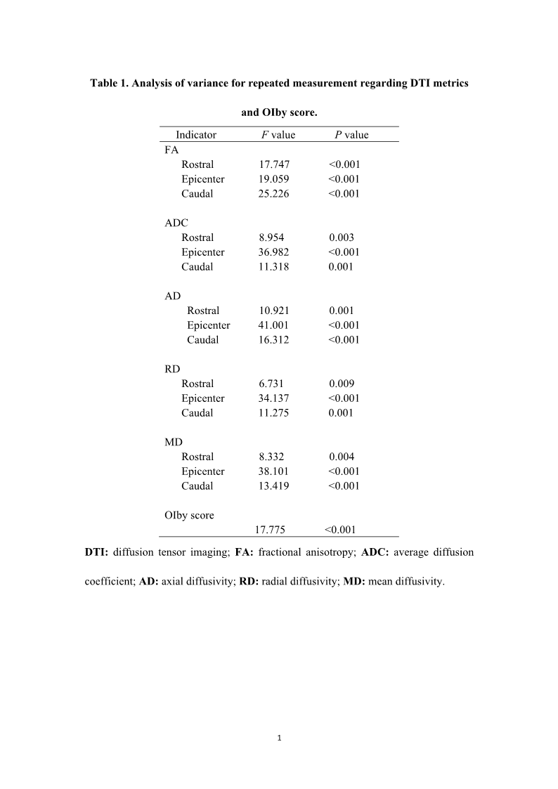
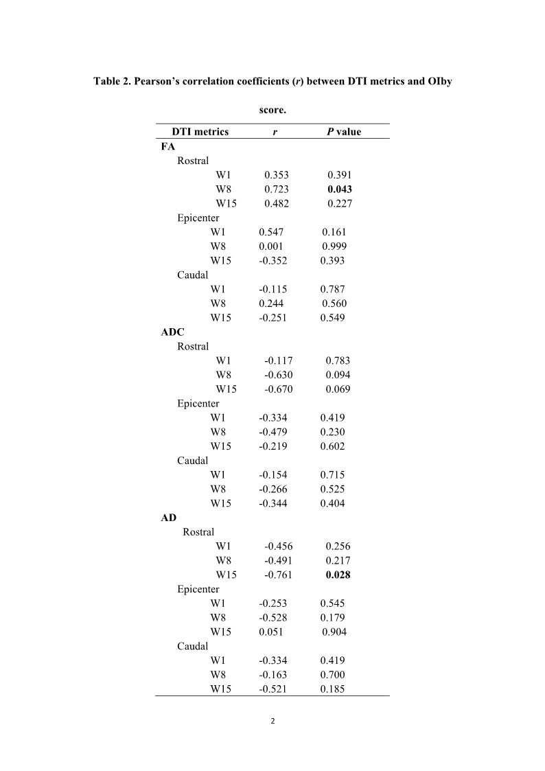
In this study, normality of continuous variables was confirmed firstly and then, they were expressed as means ± standard deviations. In terms of OIby score and radiological data collected at multiple (≥ 3) time points, analysis of variance for repeated measurement was used to test general statistical significance. Following Bonferroni correction, the paired samples t-test was performed. Pearson correlation analysis was used to detect correlation between all the radiological indicators at multiple time points and OIby score at W15. After determining a DTI parameter showing the highest correlation with the final pelvic limb locomotion, simple linear regression model was established to estimate behavioral change by using this imaging indicator as a predictor. SPSS Statistics version 22.0 (IBM Corporation, United States) was applied to perform aforementioned statistical analyses, and P<0.05 was considered statistically significant. All graphs were made with GraphPad Prism version 5.0 (GraphPad Software, United States).
Results
Motor restoration of SCI canines
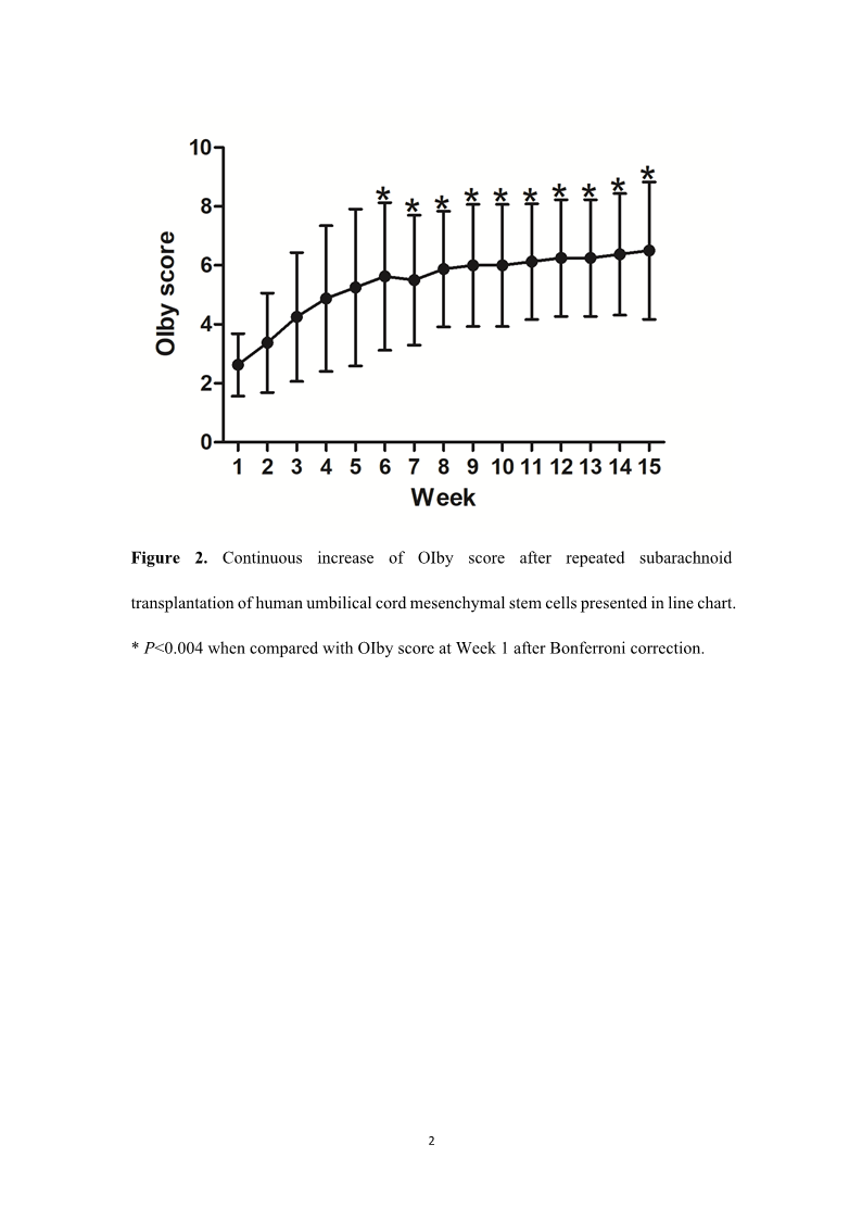
All the canines received SCI modeling and subsequent four times of subarachnoid transplantation of hUC-MSCs smoothly, and no animal died before the study endpoint. During the whole observation period, no immune rejection was found in these canines, and their OIby scores increased gradually. At W1, its mean value was 2.6, while it increased to 5.9 and 6.5 at W8 and W15, respectively. Although transient decline of OIby score could be found at W7, its overallstatistical significance was demonstrated during the 15-week observation period (Table 1). The OIby score collected at the latest ten time points showed significant improvements when compared with that at W1 (Figure 2).
Temporal changes in DTI parameters
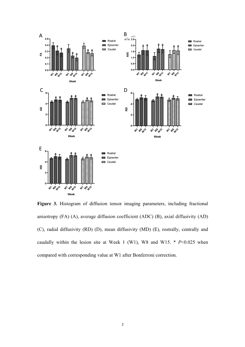
With respect to mean values of radiological metrics, FA rostrally, centrally and caudally within the lesion site revealed the decrease tendency, while all the other radiological indicators (ADC, AD, RD, MD) at these three regions had the N-shaped trend that showed initial increase at W8 and subsequent decrease at W15. All the radiological parameters were proved to have overall statistical significance (Table 1). Except the comparisons between W15 and W1 regarding rostral MD, rostral RD and caudal RD, the remaining comparisons of radiological data between W15 and W1 demonstrated statistical significance. Moreover, all the radiological parameters between W8 and W1 also showed statistical significance (Figure 3).
Correlation between DTI parameters and motor function regaining
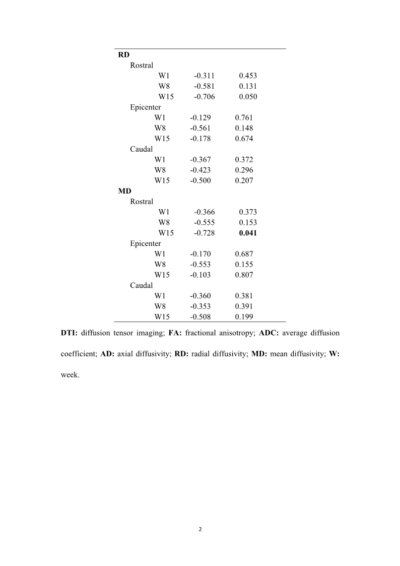
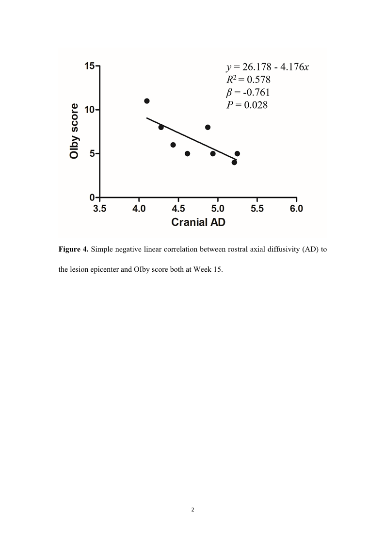
According to Pearson correlation analysis, rostral FA (r=0.723, P=0.043) at W8, as well as both cranial AD (r=-0.761, P=0.028) and MD (r=-0.728, P=0.041) at W15 were found to correlate with the final OIby score, while all the other DTI parameters had no coorelation (Table 2). Among the aforementioned radiological metrics, rostral AD at W15 had the relatively highest correlation, and then simple regression model (y=26.178-4.176x, y: OIby score, x:cranial AD value) was constructed to further uncovered a negative linear correlation between rostral AD and locomotion performance both at W15 (R2=0.578, β=-0.761, P=0.028) (Figure 4). Accordingly, it was estimated that in canine model of SCI, OIby score could increase by approximate four points when cranial AD decreased by one point at 10 weeks after four times of subarachnoid administrations of hUC-MSCs.
Discussion
This prospective study aimed to investigate the dynamic changes in DTI parameters following repeated intrathecal administrations of hUC-MSCs and their correlations with motor function recovery in canine model of SCI. Our principal findings were as follows. Firstly, repeated hUC-MSCs transplantations promoted significant and sustained improvement in locomotor function, as assessed by the Olby score. Secondly, DTI parameters (FA, ADC, AD, RD, MD) at the lesion epicenter, as well as in adjacent rostral and caudal regions exhibited significant temporal changes throughout the 15-week observation period. Thirdly, specific DTI metrics, including rostral FA at 3 weeks, as well as rostral AD and MD at 10 weeks after hUC-MSCs delivery, demonstrated significant correlations with the final behavioral outcome. Among these indicators, rostral AD at the study endpoint emerged as a potential predictive biomarker for motor recovery.
While the primary focus of this study is on correlations between DTI metrics and motor recovery in canine model of SCI, the observed functional restoration is mainly attributed to the therapeutic effects of subarachnoid administrations of hUC-MSCs. The chosen intrathecal route facilitates widespread distribution and higher engraftment of cells within the cerebrospinal fluid, allowing them to exert their effects along the spinal axis 10, 11. The therapeutic benefits of hUC-MSCs are likely mediated through powerful paracrine mechanisms rather than direct cell replacement 5, 7, 9. Specifically, the inflammatory microenvironment post-SCI can be modulated towards a more anti-inflammatory state. Moreover, hUC-MSCs can secrete a plethora of neurotrophic factors, including brain-derived neurotrophic factor, nerve growth factor, to promote neuronal survival, axonal sprouting, and remyelination of damaged axons. Furthermore, hUC-MSCs support angiogenesis to improve blood supply to the compromised tissue and modulate glial scar formation to create a more permissive environment for neurological repair 12-14.
DTI is invaluable to capturing microstructural changes distant from the injury epicenter not evident on conventional MRI T2 weighted image, and thus it enhances detection of subtle pathology in the whole spinal cord 18. Based on this advantage, both rostral and caudal DTI indicators of damaged spinal cord in the canine model were analyzed to make a more comprehensive understanding of pathophysiological change after cytotherapy. Because lesion severity is inversely related to distance from the injury epicenter 19, it is reasonable that rostral and caudal ROI were set within one vertebral body above or below the visible lesion margins based on MRI T2 weighted image. Specifically, FA provides a reliable measure of fiber quantity loss and myelin structural disruption in white matter 20,21. Although canines showed varying degrees of behavioral recovery following hUC-MSCs transplantation, the number and integrity of white matter might not be regained, leading to no increase of FA within the 15-week observation period. ADC indicates the density of nerve fibers in white matter and the diffusion of water molecules in gray matter 22,23. After SCI, it increased because of axonal disruption and demyelination, as well as enhanced anisotropic diffusion of water molecules 22,23. Even though hUC-MSCs infusion could promote motor function recovery, intra-axonal ultrastructural changes inherent of the secondary injury, such as mitochondrial accumulation, might still represent a long-lasting effect on diffusion magnitude. Hence, these pathological changes caused continued increase of ADC within a period 24. Following axonal repair, it began to decline. AD is sensitive to axonal injury and influenced by its structural integrity 25. Initially, AD increased possibly because of demyelination and vacuolation according to progression of the damage to spinal cord even if hUC-MSCs were infused, then it began to decline 19. As an indicator of myelination state and water diffusion, RD is capable of detecting white matter and gray matter pathologies 20,23. During the initial recovery phase after SCI, the loss, demyelination and fragmentation of axons, as well as water anisotropic diffusion continued, resulting in the increase of RD despite administrations of hUC-MSCs 20,23,26,27. As white matter remyelination dominated the repair procedure, it gradually decreased after W8. Similarly, MD also responds to aforementioned white matter and gray matter lesions triggered by SCI and may be used as a parameter to differentiate injury severity based on restricted diffusivity 20,23,28. It increased over the early recovery period, which might be due to long-lasting parenchymal tissue edema and disruption, as well as subsequently strengthened diffusion of water molecules 27. Moreover, continuous decrease in FA would also be influenced by increased MD. Following hUC-MSCs delivery, the local microenvironment within the damaged spinal cord improved, leading to the decrease of MD.
Our findings were similar to another two reports recruiting patients suffering from cervical SCI which demonstrated a strong correlation between AD and neurological outcomes, as well as a higher ability of MD measured one level rostrally to the lesion epicenter in predicting neurological recovery 29,30. This phenomenon may be explained by histological results that show the rostral segment of the lesioned dorsal column undergoes more severe demyelination and degeneration, while residual supraspinal input has important effect on regaining locomotion of limbs 19,20,27,31. The differences of DTI parameters observed in rostral versus epicenter or caudal damaged levels indicate ascending and descending tracts may react differently to cytotherapy probably due to a higher white-to-gray matter ratio in rostral site, and thus they provide potential indices to predict therapeutic outcomes 32. According to our findings, rostral FA at 3 weeks post-cytotherapy, as well as both cranial AD and MD at 10 weeks after hUC-MSCs administrations appeared to be most sensitive in detecting pathological changes of damaged spinal cord. Of these three parameters, rostral AD at 10 weeks after repeated intrathecal administrations of hUC-MSCs had the relatively highest correlation with the final regaining of motor function. Moreover, the change in finally measured AD rostrally to the lesion epicenter seems a feasible predictor of behavioral recovery based on simple linear regression analysis. It is supposed that rostral AD at final follow-up served as a potential quantitative imaging indicator to predict sensorimotor recovery after hUC-MSCs administrations in humans. Such DTI profiles will enable clinicians to decide whether supplementary therapeutic strategies should be implemented to enhance clinical efficacy of cytotherapy.
This study has several limitations that must be acknowledged. Firstly, the lack of a control group prevents researchers from definitively attributing all observed motor recovery and DTI changes solely to hUC-MSCs intervention, as some changes may reflect natural history of SCI repair. Further studies with a control group are essential to differentiate therapeutic effects of hUC-MSCs from spontaneous neurological recovery after SCI. Secondly, the 15-week observation period may be inappropriate to assess the long-term outcomes. Thirdly, survival, distribution and differentiation of the transplanted hUC-MSCs were not assessed through histology or in vivo tracking, which probably provide crucial mechanistic insights linking the DTI changes to cellular-level events. Fourthly, motor assessment fully relied on Olby score. Incorporating additional objective gait analysis tools, including kinematic analysis or pressure-sensitive walkways, may provide a more comprehensive evaluation. Finally, DTI measurements were only based on the whole cross-sections of spinal cord. Further studies employing tract-specific analysis may pinpoint changes in specific motor and sensory pathways, offering greater predictive specificity.
Conclusions
For the first time, our work demonstrates that in canine model of SCI receiving repeated intrathecal transplantation of hUC-MSCs, rostral FA at 3 weeks post-cytotherapy, as well as both cranial AD and MD at 10 weeks after hUC-MSCs administrations, demonstrated significant correlations with the final motor function regaining, with rostral AD emerging as a potential imaging predictor. This study provides important imaging clues for prediction of sensorimotor function restoration in humans suffering from SCI after repeated subarachnoid transplantation of hUC-MSCs.

quarterly,launched in March 2025
Editor-in-Chief: Limin Rong
Sponsor: Sun Yat-sen University
Publisher: Sun Yat-sen University Press
Co-Publisher: KeAi Communications Co., Ltd.
Edited by: Editorial Office of Journal of Brain and Spine
Address: 600 Tianhe Road, Guangzhou, 510630, China
Website: http://jbs.sypub.cn/jbs
E-mail: jbseditor@mail.sysu.edu.cn

Address:600 Tianhe Road, Guangzhou, 510630, China
Website:http://jbs.sypub.cn/jbs
E-mail:jbseditor@mail.sysu.edu.cn